✍️ Author: Dr Eleni Christoforidou
Home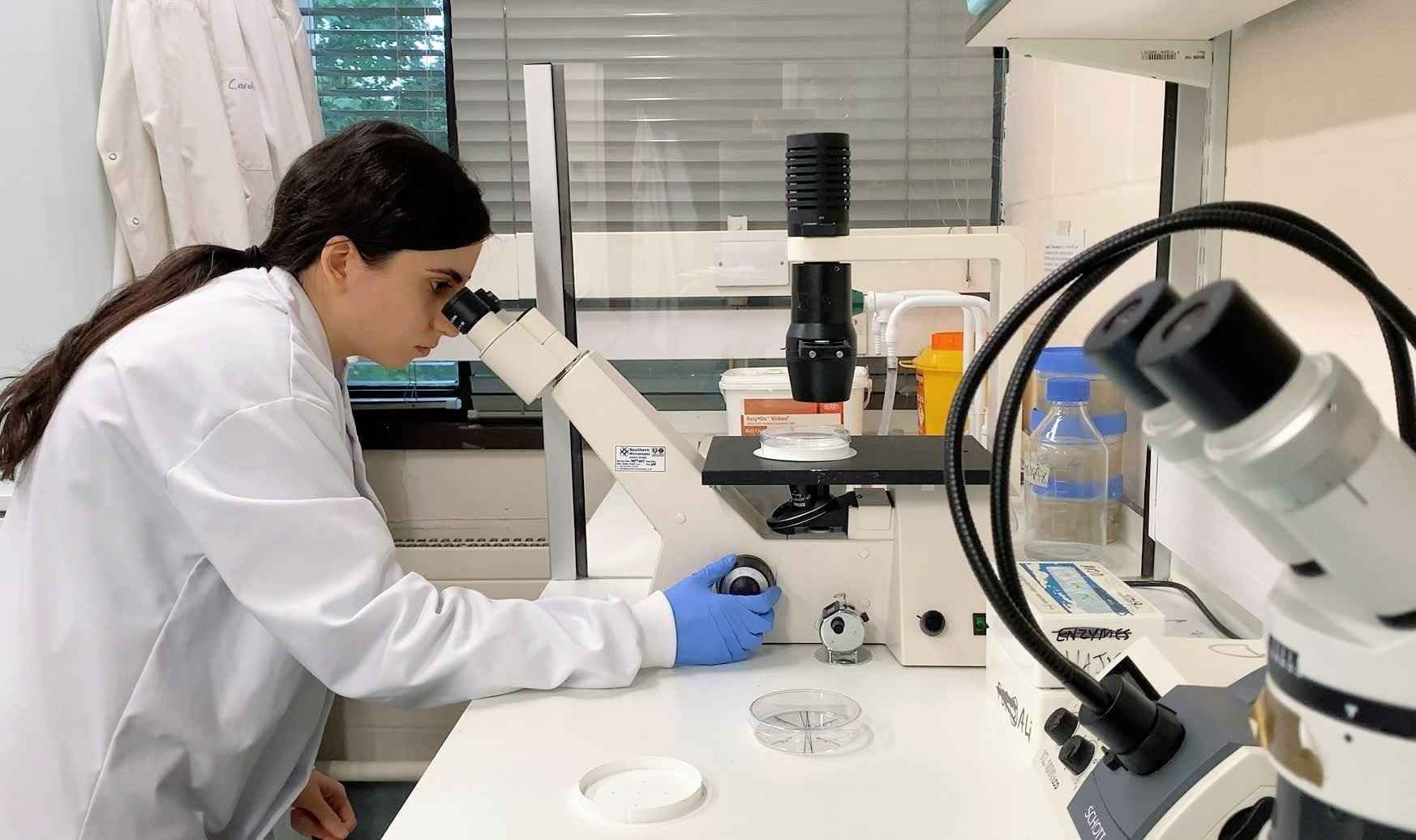
Using a phase-contrast microscope.
A Day in the Lab: Harnessing a Nobel-Winning Technique
🕒 Approximate reading time: 3 minutes
Today, our lab adventure takes us on a journey through the fascinating world of cells, leveraging the power of phase-contrast microscopy.
Phase-contrast microscopy is a groundbreaking optical microscopy technique that transforms phase shifts in light passing through a transparent specimen into brightness variations in the resultant image. Although phase shifts are not visible in themselves, they become noticeable when displayed as alterations in brightness.
When light waves traverse a medium other than a vacuum, they interact with this medium, causing changes in wave amplitude and phase, dependent on the properties of the medium. Changes in amplitude, or brightness, stem from the scattering and absorption of light, which is often wavelength-dependent, and may give rise to colours. However, without specific provisions, phase changes remain invisible since photographic equipment and the human eye are primarily sensitive to amplitude variations.
Phase-contrast microscopy brings to light numerous cellular structures that remain hidden with a conventional bright-field microscope. Previously, these structures could only be made visible through staining, a process requiring additional preparation and the inevitable death of the cells. The advent of the phase-contrast microscope enabled biologists to study living cells in real-time, observing their proliferation through cell division. It remains one of the few methods that allow for the quantification of cellular structure and components without resorting to fluorescence.
This technique's significance and potential were so groundbreaking that its inventor, Frits Zernike, was awarded the Nobel Prize in Physics in 1953, not long after its inception in the early 1930s. Join us next time as we continue to delve into the world of cells, unearthing more of their secrets through the lens of our powerful microscopes.