✍️ Author: Dr Eleni Christoforidou
Home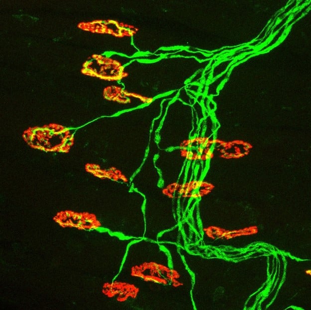
The neuromuscular junctions are shown in red and the neurons are shown in green.
A Day in the Lab: Microscopic Exploration of Muscle Biopsies
🕒 Approximate reading time: 2 minutes
Today, our lab adventure involves muscle biopsies, specifically those from the feet. After undergoing an immunostaining process with fluorescent antibodies, these minute biopsies are mounted on microscope slides. The aim of this process is to highlight the neuromuscular junctions, where a neuron interacts with a muscle, instructing it to either contract or relax.
Timelapse: Mounting muscle biopsies onto microscope slides.
Post-immunostaining, these muscle samples are situated onto microscope slides, preparing them for examination using confocal microscopy. The purpose of this analysis is to ascertain whether a genetic anomaly in the motor neurons impacts the development of these neuromuscular junctions.
The image below offers an example of how these samples appear when scrutinised under the microscope.